Embark on an engaging exploration of human anatomy through art-labeling activity the anatomy of the gallbladder and bile ducts, an innovative approach that fosters a deeper comprehension of the intricacies of the biliary system. This activity seamlessly blends artistic expression with scientific inquiry, inviting students to actively engage with the anatomical structures of the gallbladder and bile ducts.
The activity revolves around a visually appealing representation of the anatomical structures, providing a tangible canvas for students to label and identify the various components. Through this hands-on experience, they gain a comprehensive understanding of the location, shape, and interconnections of these vital organs.
Anatomy of the Gallbladder
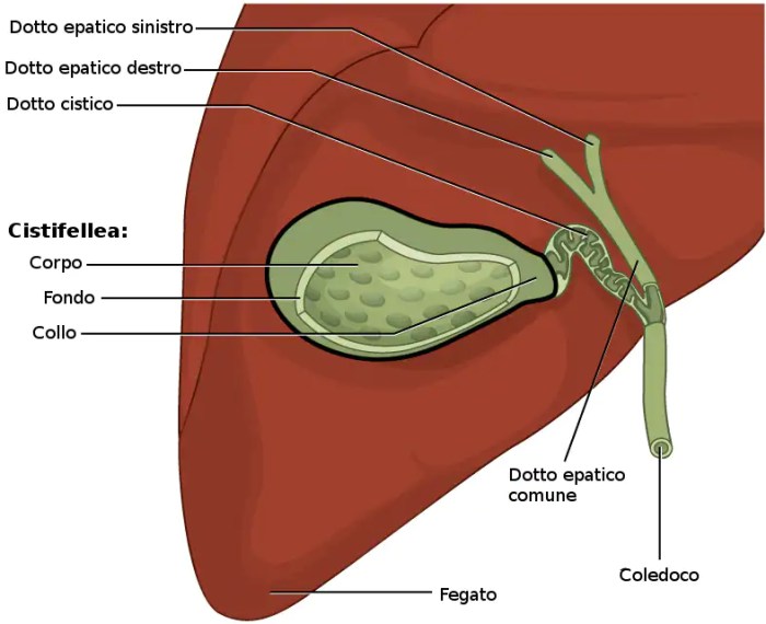
The gallbladder is a small, pear-shaped organ located on the underside of the liver. It stores and concentrates bile, a fluid produced by the liver that aids in the digestion of fats. The gallbladder has a thin wall consisting of three layers: the serosa, muscularis, and mucosa.
The serosa is the outermost layer and is continuous with the peritoneum, the lining of the abdominal cavity. The muscularis is the middle layer and is responsible for contracting the gallbladder to expel bile. The mucosa is the innermost layer and is lined with epithelial cells that secrete mucus.
The gallbladder is connected to the liver by the cystic duct and to the duodenum by the common bile duct.
Anatomy of the Bile Ducts

The bile ducts are a system of tubes that transport bile from the liver to the duodenum. The main bile ducts are the common bile duct, hepatic ducts, and cystic duct. The common bile duct is formed by the union of the right and left hepatic ducts and carries bile from the liver to the duodenum.
The hepatic ducts are the branches of the common bile duct that carry bile from the liver to the common bile duct. The cystic duct is the duct that connects the gallbladder to the common bile duct. The sphincter of Oddi is a muscle that surrounds the opening of the common bile duct into the duodenum and regulates the flow of bile into the duodenum.
Art-Labeling Activity: Gallbladder and Bile Ducts
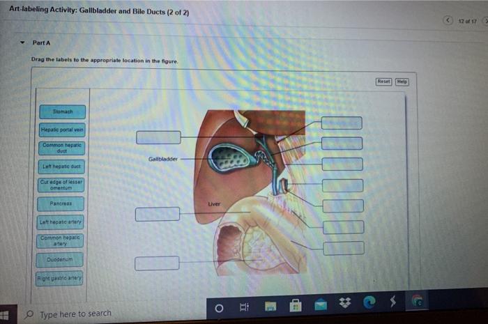
The art-labeling activity is an interactive way for students to learn about the anatomy of the gallbladder and bile ducts. The activity can be used as a review of the material or as a way to introduce the material. The activity can be completed individually or in groups.To
complete the activity, students will need a diagram of the gallbladder and bile ducts. Students will then label the different structures on the diagram. The structures that students will label include the gallbladder, liver, common bile duct, hepatic ducts, cystic duct, and sphincter of Oddi.
Applications of Art-Labeling Activity

The art-labeling activity is a valuable tool for teaching anatomy. The activity helps students to visualize the anatomy of the gallbladder and bile ducts and to learn the names of the different structures. The activity also helps students to develop their spatial reasoning skills.The
art-labeling activity can be used in a variety of educational settings. The activity can be used in a traditional classroom setting, in a laboratory setting, or in an online setting. The activity can also be used as a homework assignment or as a review activity.
Extensions and Modifications: Art-labeling Activity The Anatomy Of The Gallbladder And Bile Ducts

The art-labeling activity can be extended or modified to meet the needs of different learners. For example, the activity can be made more challenging by adding more structures to the diagram. The activity can also be modified to make it more accessible for students with disabilities.
For example, the activity can be completed using a computer or a tablet.
Essential FAQs
What are the benefits of using art-labeling activities in anatomy education?
Art-labeling activities offer numerous benefits in anatomy education. They enhance visual learning, promote active engagement, improve spatial reasoning, and foster a deeper understanding of anatomical structures and their relationships.
How can I incorporate art-labeling activities into my teaching environment?
Incorporating art-labeling activities into your teaching environment is straightforward. You can use illustrations, diagrams, or even create your own visual representations of anatomical structures. Provide clear instructions for students to follow while labeling, ensuring they engage actively with the material.
Can art-labeling activities be adapted for different grade levels or educational settings?
Absolutely. Art-labeling activities can be tailored to suit various grade levels and educational settings. By adjusting the complexity of the visual representations and the level of detail required for labeling, you can make these activities accessible to students of all levels.